
What is Hip Dysplasia?
Hip dysplasia is a medical condition that involves abnormal development or formation of the hip joint. The hip joint is a ball-and-socket joint where the head of the femur (thigh bone) fits into the acetabulum (socket) of the pelvis. In individuals with hip dysplasia, there is a mismatch between the size and shape of the femoral head and the acetabulum, which can lead to instability, poor alignment, and potential damage to the joint surfaces.
Hip dysplasia can develop in infants, adolescents, and adults. There are two main types of hip dysplasia:
- Developmental Dysplasia of the Hip (DDH): This condition typically presents in infants and young children. It occurs when the hip joint fails to develop properly, resulting in an unstable hip joint. In mild cases, the femoral head might be loosely held within the acetabulum, while in severe cases, the femoral head might be entirely dislocated from the socket.
- Acetabular Dysplasia: This form of hip dysplasia often becomes evident during adolescence or early adulthood. It involves a shallow or underdeveloped acetabulum, which doesn't provide sufficient coverage for the femoral head. This can lead to hip instability and increased risk of hip labral tears, cartilage damage, and early onset of osteoarthritis.
The causes of hip dysplasia can be multifactorial and may involve a combination of genetic predisposition, abnormal fetal positioning in the womb, and other environmental factors. Risk factors for hip dysplasia include being female (as it is more common in females), family history of the condition, and breech positioning during pregnancy (where the baby's buttocks or feet are positioned to be delivered first).
Treatment for hip dysplasia varies depending on the severity of the condition and the age of the individual. In infants, methods such as Pavlik harness or spica casting might be used to hold the hip joint in a stable position, allowing for proper development. In older children and adults, treatment might involve physical therapy, bracing, or, in more severe cases, surgical interventions like hip osteotomy (reshaping the bones) or hip replacement.
Early detection and appropriate treatment are crucial for managing hip dysplasia and minimizing the risk of long-term joint problems. Regular checkups with healthcare providers, especially for infants and young children, can aid in the timely diagnosis and management of this condition.
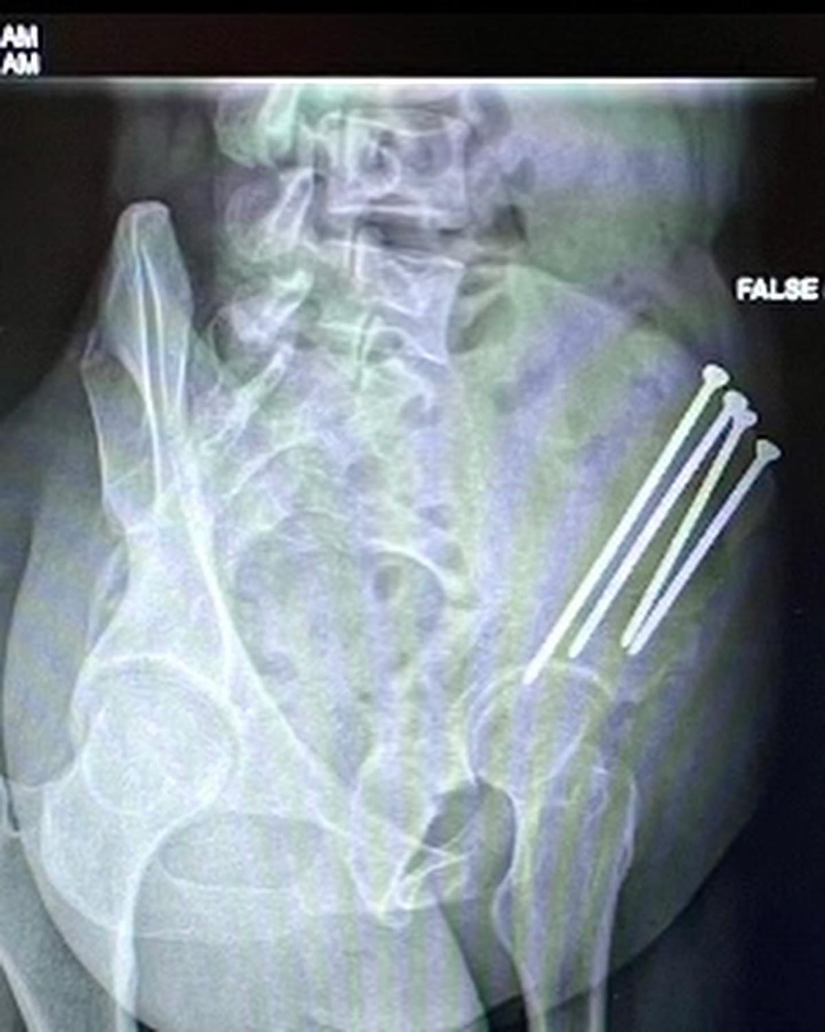
A periacetabular osteotomy (PAO) is a surgical procedure performed to treat hip dysplasia, specifically addressing acetabular dysplasia. During a periacetabular osteotomy, the surgeon repositions the acetabulum to improve its coverage of the femoral head (the ball part of the hip joint). This is done by making cuts in the pelvic bone around the acetabulum and then repositioning the acetabulum to provide better alignment and stability to the hip joint. The procedure aims to achieve better congruence between the femoral head and the acetabulum, which can help prevent further joint damage and reduce the risk of arthritis.
Here are the general steps of a periacetabular osteotomy:
- Preparation: The patient is placed under general anesthesia or spinal anesthesia with IV sedation. Surgical tools, imaging equipment (such as fluoroscopy or intraoperative X-rays), and monitoring devices are set up.
- Incisions: The surgeon makes an incision around the hip area, generally in the bikini line, to access the pelvic bone and the acetabulum.
- Bone Cuts: The surgeon carefully makes cuts in the pelvic bone around the acetabulum, creating several bone segments.
- Repositioning: The acetabular bone segments are repositioned to improve its coverage of the femoral head. The goal is to achieve a more stable and congruent hip joint.
- Fixation: Special surgical instruments like screws, plates, or other devices are used to secure the repositioned bone segments in their new alignment. These fixation devices help stabilize the bone as it heals.
- Closure: The incisions are closed using sutures or staples, and the wound is dressed.
Recovery after a periacetabular osteotomy can take several months, up to a year, and it involves a period of restricted weight-bearing and physical therapy to help the patient regain strength, mobility, and flexibility in the hip joint. The success of the procedure largely depends on the patient's commitment to following postoperative guidelines and rehabilitation.
Periacetabular osteotomy is a complex procedure that requires specialized training and experience in hip surgery. It is generally considered for younger patients who have acetabular dysplasia and are at risk of developing hip joint problems later in life. The decision to undergo a PAO is typically made based on the individual's specific condition, age, lifestyle, and overall health. It's important for patients to consult with a qualified orthopedic hip surgeon with a specialty in hip preservation, to determine the most appropriate treatment approach for their hip dysplasia.